Cell biology
Published projects:
→ Simultaneous dual-color 3D STED microscopy
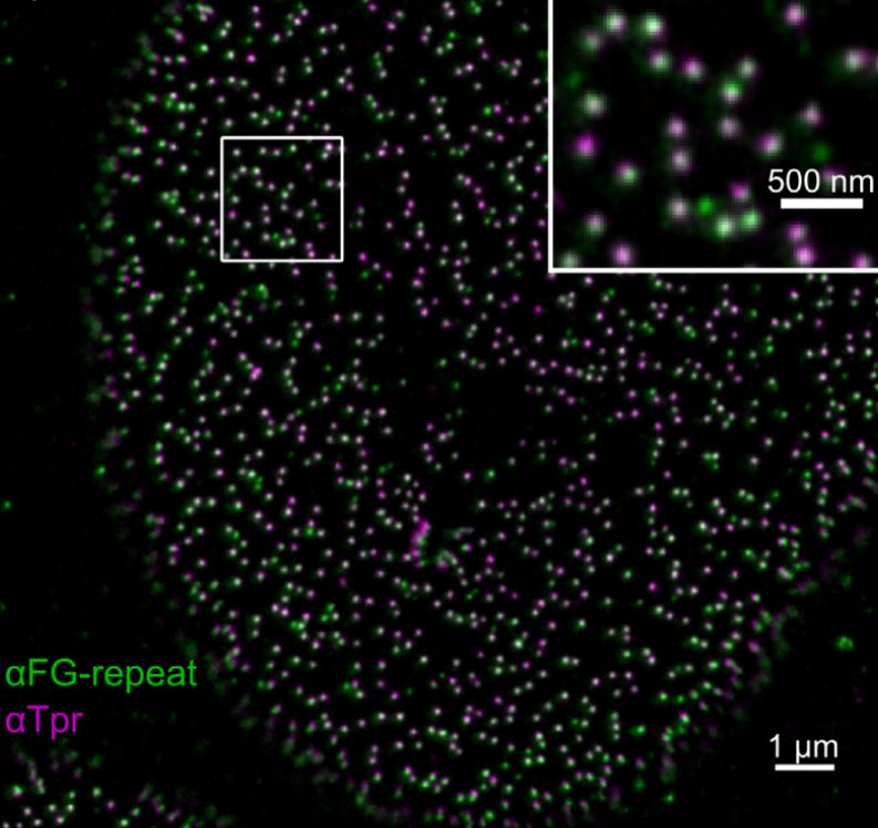
We describe the design and implementation of a stimulated emission depletion (STED) microscope which allows simultaneous three-dimensional super-resolution imaging in two colors. A super-continuum laser source is used to provide all spectral bands necessary for excitation and efficient depletion to achieve a lateral and axial resolution of ~35 nm and ~90 nm respectively. We characterize the systems' performance by imaging colloidal particles and single fluorescent molecules. Its biological applicability is demonstrated by dual-color imaging of nuclear pore complexes and of DNA replication sites in mammalian cells.
DOI: 10.1364/OE.22.007028
→ Cyclophilin-Facilitated Membrane Trabslocation as Pharmacological Target to Prevent Intoxication of Mammalian Cells by Binary Clostridial Actin ADP-Ribosylated Toxins
Clostridiumbotulinum C2 toxin, Clostridiumperfringens iota toxin and Clostridiumdifficile CDT belong to the family of binary actin ADP-ribosylating toxins and are composed of a binding/translocation component and a separate enzyme component. The enzyme components ADP-ribosylate G-actin in the cytosol of target cells resulting in depolymerization of F-actin, cell rounding and cell death. The binding/translocation components bind to their cell receptors and form complexes with the respective enzyme components. After receptor-mediated endocytosis, the binding/translocation components form pores in membranes of acidified endosomes and the enzyme components translocate through these pores into the cytosol. This step is facilitated by the host cell chaperone heat shock protein 90 and peptidyl-prolyl cis/trans isomerases including cyclophilin A. Here, we demonstrate that a large isoform of cyclophilin A, the multi-domain enzyme cyclophilin 40 (Cyp40), binds to the enzyme components C2I, Ia and CDTa in vitro. Isothermal titration calorimetry revealed a direct binding to C2I with a calculated affinity of 101 nM and to Ia with an affinity of 1.01 μM. Closer investigation for the prototypic C2I revealed that binding to Cyp40 did not depend on its ADP-ribosyltransferase activity but was stronger for unfolded C2I. The interaction of C2I with Cyp40 was also demonstrated in lysates from C2-treated cells by pull-down. Treatment of cells with a non-immunosuppressive cyclosporine A derivative, which still binds to and inhibits the peptidyl-prolyl cis/trans isomerase activity of cyclophilins, protected cells from intoxication with C2, iota and CDT toxins, offering an attractive approach for development of novel therapeutic strategies against binary actin ADP-ribosylating toxins.
DOI:10.1016/j.jmb.2014.07.013
→ A fast analysis method to quantify nanoparticle uptake on a single cell level
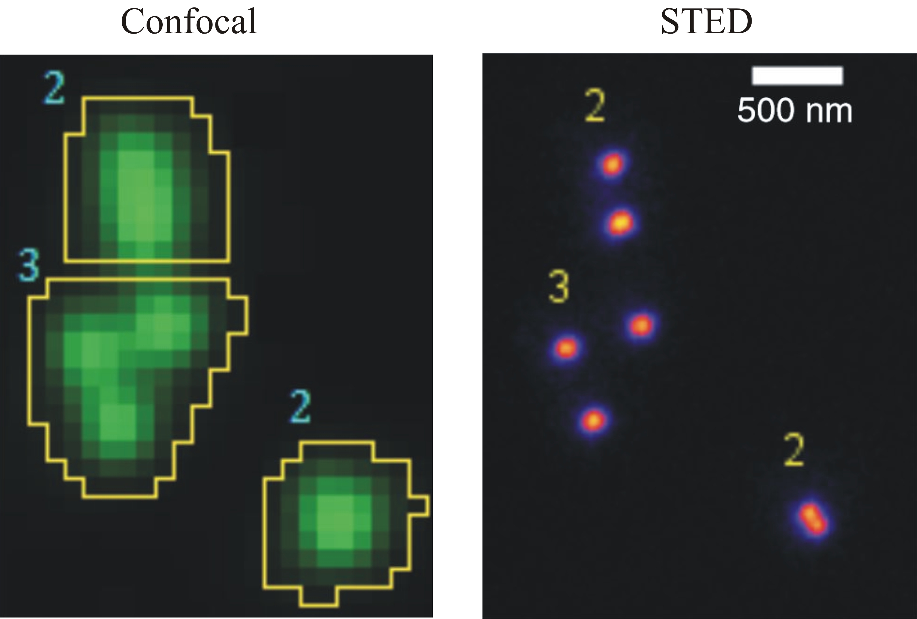
This study examines the absolute quantification of particle uptake into cells. Methods: We developed a novel method to analyze stacks of confocal fluorescence images of single cells interacting with nanoand micro-particles. Particle_in_Cell-3D is a freely available ImageJ macro. During the image analysis routine, single cells are reconstructed in 3D and split into two volumes - intracellular and the membrane region. Next, particles are localized and color-coded accordingly. The mean intensity of single particles, measured in calibration experiments, is used to determine the absolute number of particles.
Results: Particle_in_Cell-3D was successfully applied to measure the uptake of 80-nm mesoporous silica nanoparticles into HeLa cells. Furthermore, it was used to quantify the absolute number of 100-nm polystyrene nanoparticles forming agglomerates of up to five particles; the accuracy of these results was confirmed by super-resolution, stimulated emission depletion microscopy. Conclusion: Particle_in_Cell-3D is a fast and accurate method that allows the quantification of particle uptake into cells.
http://dx.doi.org/10.2217/NNM.12.178