Overview
Spinning-disk confocal microscopy is applied using different fluorescence tagging strategies.
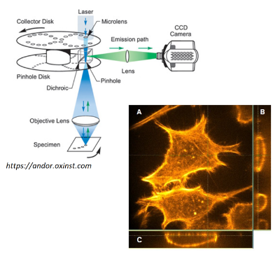
The actin cytoskeleton of fixed HeLa cells is stained by rhodamine phalloidin, which acts like a highly specific fluorescent antibody.
Intracellular calcium signals following receptor stimulation are studied using Fluo4, a cell permeant calcium indicator dye.
Dynamics in the mitochondrial network is studied using mt-Eos, a photoactivatable protein which carries a mitochondrial targeting signal.
![[Translate to english:] Button Overview Experiments](/fileadmin/website_uni_ulm/nawi.physik/dateien/data/biophyscis_lab/button_overview_experiments_350e.png)