Instrument development
Current research:
We develop a reflected light sheet microscope for convenient imaging of single florescent proteins in living zebrafish embryos.
We develop a high sensitivity single molecule force spectroscopy instrument to study the mechano-kinetic properties of biomolecules and their interactions.
Published projects:
Quantum Optical Microphone in the Audio Band
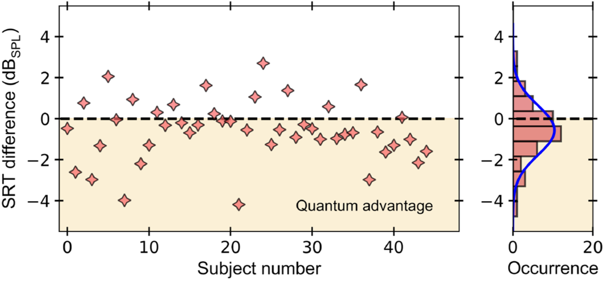
The ability to perform high-precision optical measurements is paramount in science and engineering. Laser interferometry enables interaction-free sensing with a precision ultimately limited by shot noise. Quantum optical sensors can surpass this limit but single- or multiphoton schemes are challenged by low experimental sampling rates, while squeezed-light approaches require complex optical setups and sophisticated time gating. Here, we introduce a simple method that infers optical phase shifts through standard intensity measurements while still maintaining the quantum advantage in the measurement precision. Capitalizing on the robustness and high sampling rates of our device, we implement a quantum optical microphone in the audio band. Its performance is benchmarked against a classical laser microphone in a standardized medically approved speech-recognition test on 45 subjects. We find that quantum-recorded words improve the speech-recognition threshold by −0.57dBSPL, thus making the quantum advantage audible. Not only do these results open the door toward applications in quantum nonlinear interferometry but they also show that quantum phenomena can be experienced by humans.
doi.org/10.1103/PRXQuantum.3.020358
→ Reflected light sheet microscopy
Single-molecule imaging of transcription factor binding to DNA in live mammalian cells
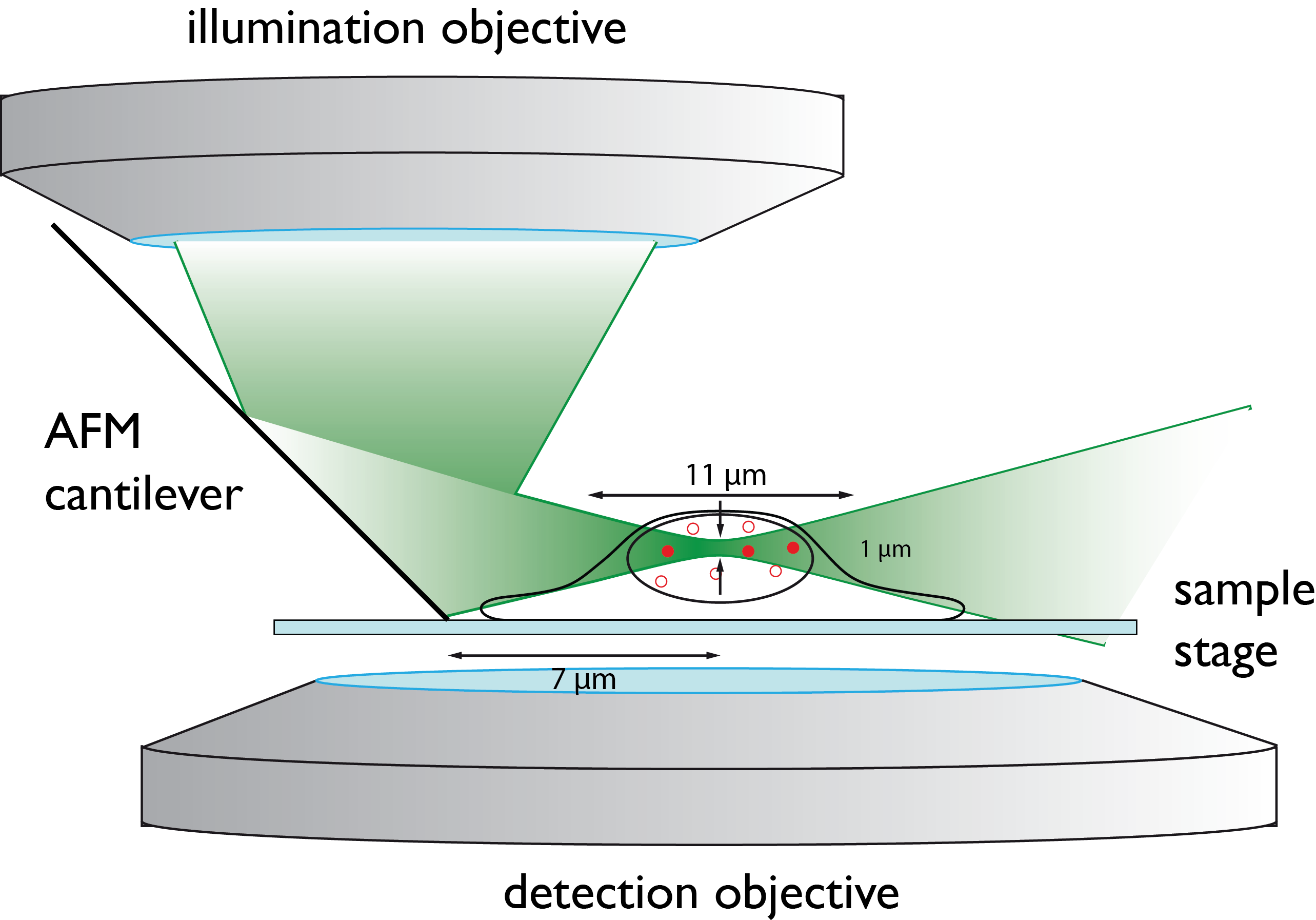
Imaging single fluorescent proteins in living mammalian cells is challenged by out-of-focus fluorescence excitation. To reduce out-of-focus fluorescence we developed reflected light-sheet microscopy (RLSM), a fluorescence microscopy method allowing selective plane illumination throughout the nuclei of living mammalian cells. A thin light sheet parallel to the imaging plane and close to the sample surface is generated by reflecting an elliptical laser beam incident from the top by 90° with a small mirror. The thin light sheet allows for an increased signal-to-background ratio superior to that in previous illumination schemes and enables imaging of single fluorescent proteins with up to 100-HZ time resolution.
doi: 10.1038/nmeth.2411