
Core Facility Manager
Dr. Christian Bökel
Tel: 0731 50 33701
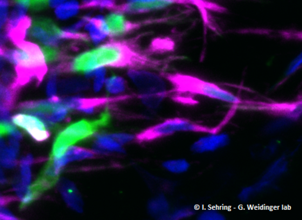
The ULMTeC Core Facility Light Microscopy can be accessed by all scientists of the Medical Faculty and Ulm University and offers a wide spectrum of optical microscopy approaches. Our main technique, confocal microscopy, is based on the pixelwise scanning with illumination lasers to generate virtual 2D optical sections through 3D objects.
The core facility primarily provides training that allows users to later operate our microscopes independently. In addition, we offer support with the evaluation and interpretation of the image data, in particular concerning quantitative and statistical image analysis. Due to our long standing expertise we can also offer help with the design of imaging based experiments and the development of bespoke experimental protocols.
QuickLinks
Equipment

Leica TCS SP8
- Inverse confocal microscope.
- Equipped with sensitive hybrid detectors.
- Optimized for quantiative fluorescence imaging.
- Equipped with a stage incubator for live cell imaging.
- Laser lines 405, 488, 561 and 633 nm.

Zeiss LSM 710
- Inverse confocal microscope
- Equipped with TCSPC detectors and a Spectra Physics MaiTai IR-Laser for pulsed excitation
- Optimized for time correlated single photon counting applications such as FLIM (fluorescence lifetime imaging) or FLIM/FRET, e.g. for metabolic imaging
- Multiphoton and SHG (second harmonic generation) - microscopy
- Equipped with incubators for live cell imaging
- Laser lines 405, 488, 561 and 633 nm.
Services
List of services
Facility services include:
- Standard and multiphoton confocal microscopy
- Live cell imaging
- Time resolved techniques such as FLIM, FLIM/FRET or FCCS
- Expertise in experiment design, staining protocols, subcellular imaging, …
- Support with quantitative image analysis and image statistics
- Hands on support for complex imaging tasks

Slide scanner VS200 from Evident
For project and application questions, please contact directly Paul Lopatta from IMOS.
- Scanning of histological sections with loading positions for 200 slides
- Brightfield, fluorescence and polarized light measurements
- SpectraSplit®7 filter set for applications with average excitation at 375, 435, 490, 545, 590, 650, and 740nm
- Variety of installed lenses for very fast and/or high-resolution applications:
- PLN2X lens (NA 0.06)
- UPLXAPO4X lens (NA 0.16)
- UPLXAPO10X lens (NA 0.4)
- UPLXAPO20X lens (NA 0.8)
- UPLXAPO100XO lens (NA 1.45)