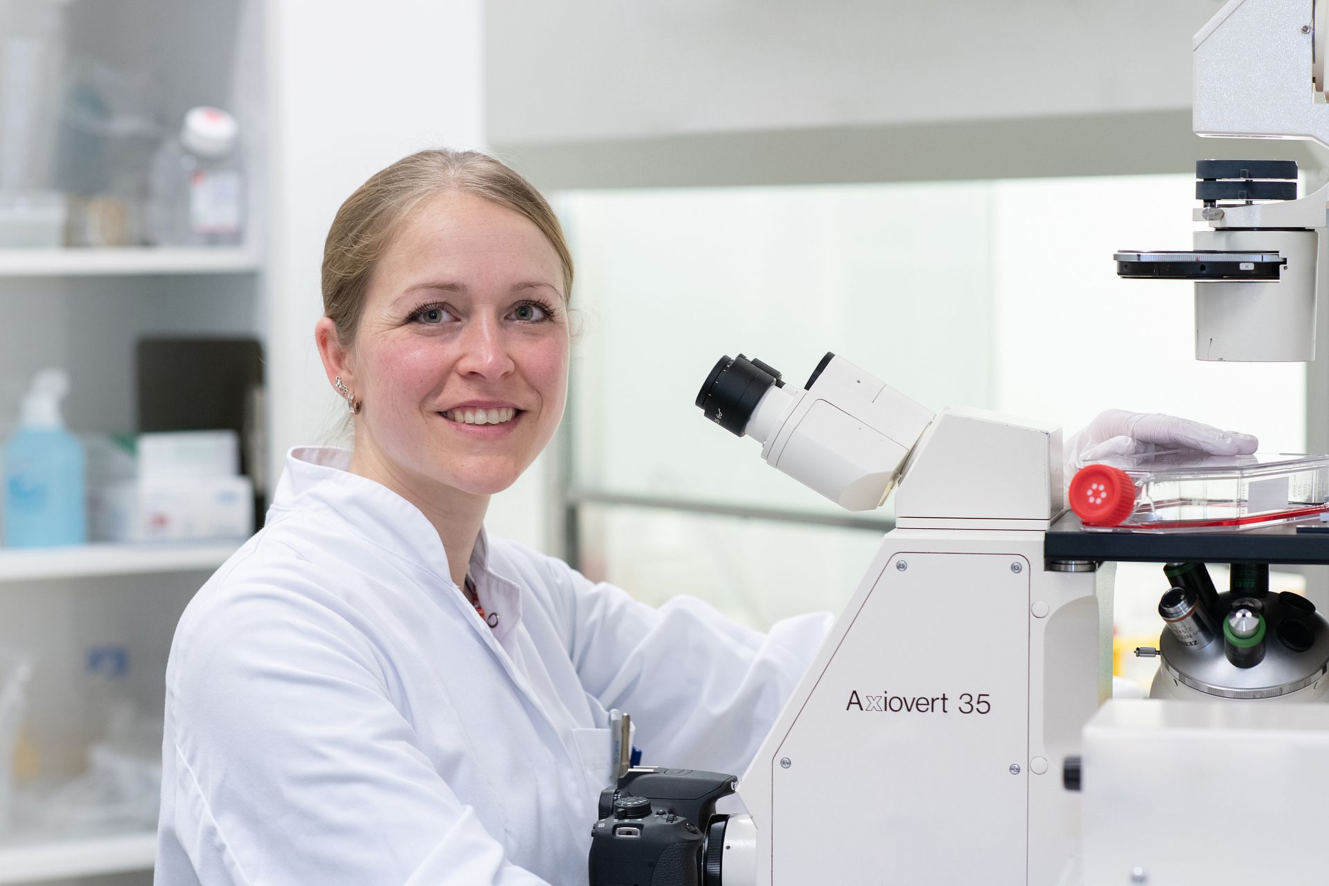
Dr. Jana Riegger-Koch
Dr. Jana Riegger-Koch is a Margarete von Wrangell-Fellow and junior group leader at the Division for Biochemistry of Joint and Connective Tissue Diseases (head: Prof. Dr. med. Rolf Brenner). Her research mainly focuses on the molecular pathomechanisms of posttraumatic osteoarthritis (PTOA).
Traumatic joint injuries are known as the main risk factor of PTOA – a special form of OA, predominantly affecting patients who are active in sports and often considerably younger than average OA patients. The pathogenetic processes include oxidative stress, (regulated) cell death, release of damage-associated molecular patterns (DAMPs) and catabolic enzymes, as well as synovial inflammation. Altogether excessive biomechanical stress and posttraumatic conditions (DAMPs, ROS, and cytokine release) result in phenotypical alterations of surviving chondrocytes. Instead of maintaining the homeostasis of the extracellular matrix and repairing the injured cartilage, dysfunctional chondrocytes promote ongoing degradation of the tissue. Although cartilage is considered as a non-regenerative tissue, there is increasing evidence of intrinsic cartilage repair by injury-responsive chondrogenic progenitor cells.
Special focus of Jana`s group is currently placed on stress-induced premature senescence as well as the contribution of complement activation products, in particular anaphylatoxins and the terminal complement complex, as potential effectors in cartilage degeneration. The projects not only aim on the clarification of the underlying pathomechanisms of PTOA but also the development of novel pharmacological and cell-based treatment strategies. To study the posttraumatic behavior and fate of surviving chondrocytes, the research group established a special human ex vivo cartilage trauma model and uses isolated human chondrocytes as well as chondrogenic progenitor cells.
Jana is funded by the DFG and is member of the CRC1149.