Single-Molecule Localization Microscopy (SMLM)
In our lab we use the single-molecule imaging techniques of STORM (stochastic optical reconstruction microscopy) and PALM (fluorescence photoactivated localization microscopy) to get insight into biophysical and biological questions.
The working principle is as follows:
Due to the diffraction of light the resolution of normal fluorescence microscopy is limited to about half the wavelength according to Abbe’s formula. This means that two nearby fluorophores can’t be resolved separately if their distance is below around 250 nm. To circumvent this basic limit one has to reach out to the time domain to gather additional spatial information. In other words: if the two nearby (i.e. their distance is well below 250 nm) fluorophores emit their fluorescence light one after the other, one can determine the center positions of the individually recorded point spread functions with high precision. Afterwards the original distribution of the fluorophores is reconstructed on the basis of the individual localizations. In this way it is possible to discern close-by positions with high precision (down to about 10 nm) using visible light even though the diffraction of light still occurs.
On the one hand we focus on building the microscope with special care taken to the specific needs of these imaging methods. Further on the homebuilt setup has the advantage of being highly adaptable to upcoming variations in illumination and detection schemes. On the other hand we apply the techniques to various research fields such as the investigation of higher order chromatin structures in vitro and protein (co-)localization in yeast or mammalian cells.
Recommended literature:
| - | Rust, M. J., Bates, M. and Zhuang, X. Nat. Methods (2006), 3, 793–795 |
| - | Betzig, E., Patterson, G. H., Sougrat, R., Lindwasser, O. W., Olenych, S., Bonifacino, J.S., Davidson, M.W., Lippincott-Schwartz, J. and Hess, H.F. Science (2006), 313, 1642–1645 |
| - | Hess, S. T., Girirajan, T. P. and Mason, M. D. Biophys. J. (2006), 91, 4258– 4272 |
Published Project:
Super-Resolution Microscopy Reveals Presynaptic Localization of the ALS/FTD Related Protein FUS in Hippocampal Neurons
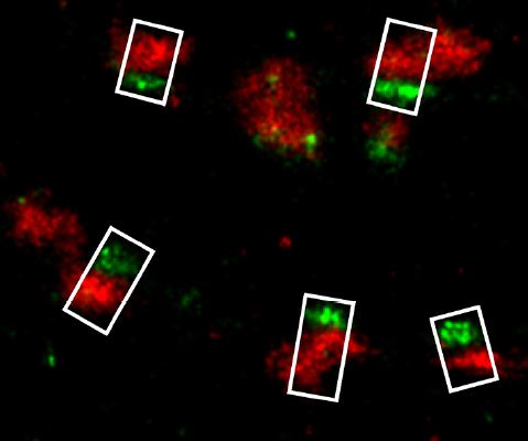
Fused in Sarcoma (FUS) is a multifunctional RNA-/DNA-binding protein, which is involved in the pathogenesis of the neurodegenerative disorders amyotrophic lateral sclerosis (ALS) and frontotemporal dementia (FTD). A common hallmark of these disorders is the abnormal accumulation of mutated FUS protein in the cytoplasm. Under normal conditions FUS is confined to the nuclear compartment, in neurons, however, additional somatodendritic localization can be observed. In this study, we carefully analyzed the subcellular localization of endogenous FUS at synaptic sites of hippocampal neurons which are among the most affected cell types in FTD with FUS pathology. We could confirm a strong nuclear localization of FUS as well as its prominent and widespread neuronal expression throughout the adult and developing rat brain, particularly in the hippocampus, the cerebellum and the outer layers of the cortex. Intriguingly, FUS was also consistently observed at synaptic sites as detected by neuronal subcellular fractionation as well as by immunolabeling. To define a pre- and/or postsynaptic localization of FUS, we employed super-resolution fluorescence localization microscopy. FUS was found to be localized within the axon terminal in close proximity to the presynaptic vesicle protein Synaptophysin1 and adjacent to the active zone protein Bassoon, but well separated from the postsynaptic protein PSD-95. Having shown the presynaptic localization of FUS in the nervous system, a novel extranuclear role of FUS at neuronal contact sites has to be considered. Since there is growing evidence that local presynaptic translation might also be an important mechanism for plasticity, FUS – like the fragile X mental retardation protein FMRP – might act as one of the presynaptic RNA-binding proteins regulating this machinery. Our observation of presynaptic FUS should foster further investigations to determine its role in neurodegenerative diseases such as ALS and FTD.
Front. Cell. Neurosci., 2016, 9:496.
doi: 10.3389/fncel.2015.00496