Publikationen 2016
Deficiency of RITA results in multiple mitotic defects by affecting microtubule dynamics

K. Steinhäuser, P. Klöble, N.N. Kreis, A. Ritter, A. Friemel, S. Roth, J. M. Reichel, J. Michaelis, M. A. Rieger, F. Louwen, F. Oswald and J. Yuan
Deregulation of mitotic microtubule (MT) dynamics results in defective spindle assembly and chromosome missegregation, leading further to chromosome instability, a hallmark of tumor cells. RBP-J interacting and tubulin-associated protein (RITA) has been identified as a negative regulator of the Notch signaling pathway. Intriguingly, deregulated RITA is involved in primary hepatocellular carcinoma and other malignant entities. We were interested in the potential molecular mechanisms behind its involvement. We show here that RITA binds to tubulin and localizes to various mitotic MT structures. RITA coats MTs and affects their structures in vitro as well as in vivo. Tumor cell lines deficient of RITA display increased acetylated α-tubulin, enhanced MT stability and reduced MT dynamics, accompanied by multiple mitotic defects, including chromosome misalignment and segregation errors. Re-expression of wild-type RITA, but not RITA Δtub ineffectively binding to tubulin, restores the phenotypes, suggesting that the role of RITA in MT modulation is mediated via its interaction with tubulin. Mechanistically, RITA interacts with tubulin/histone deacetylase 6 (HDAC6) and its suppression decreases the binding of the deacetylase HDAC6 to tubulin/MTs. Furthermore, the mitotic defects and increased MT stability are also observed in RITA−/− mouse embryonic fibroblasts. RITA has thus a novel role in modulating MT dynamics and its deregulation results in erroneous chromosome segregation, one of the major reasons for chromosome instability in tumor cells.
Oncogene [Epub ahead of print]
doi:10.1038/onc.2016.372
Energy-based scheme for reconstruction of piecewise constant signals observed in the movement of molecular machines
J. Rosskopf, K. Paul-Yuan, M. B. Plenio and J. Michaelis
Analyzing the physical and chemical properties of single DNA-based molecular machines such as polymerases and helicases requires to track stepping motion on the length scale of base pairs. Although high-resolution instruments have been developed that are capable of reaching that limit, individual steps are oftentimes hidden by experimental noise which complicates data processing. Here we present an effective two-step algorithm which detects steps in a high-bandwidth signal by minimizing an energy-based model (energy-based step finder, EBS). First, an efficient convex denoising scheme is applied which allows compression to tuples of amplitudes and plateau lengths. Second, a combinatorial clustering algorithm formulated on a graph is used to assign steps to the tuple data while accounting for prior information. Performance of the algorithm was tested on Poissonian stepping data simulated based on published kinetics data of RNA polymerase II (pol II). Comparison to existing step-finding methods shows that EBS is superior in speed while providing competitive step-detection results, especially in challenging situations. Moreover, the capability to detect backtracked intervals in experimental data of pol II as well as to detect stepping behavior of the Phi29 DNA packaging motor is demonstrated.
Phys. Rev. E, 2016, 94, 022421
Doi: 10.1103/PhysRevE.94.022421


Bottom-Up Fabrication of Nanopatterned Polymers on DNA Origami by In Situ Atom-Transfer Radical Polymerization
Y. Tokura, Y. Jiang, A. Welle, M. H.Stenzel, K. M. Krzemien, J. Michaelis, R. Berger, C. Barner-Kowollik, Y. Wu and T. Weil
Bottom-up strategies to fabricate patterned polymers at the nanoscale represent an emerging field in the development of advanced nanodevices, such as biosensors, nanofluidics, and nanophotonics. DNA origami techniques provide access to distinct architectures of various sizes and shapes and present manifold opportunities for functionalization at the nanoscale with the highest precision. Herein, we conduct in situ atom-transfer radical polymerization (ATRP) on DNA origami, yielding differently nanopatterned polymers of various heights. After cross-linking, the grafted polymeric nanostructures can even stably exist in solution without the DNA origami template. This straightforward approach allows for the fabrication of patterned polymers with low nanometer resolution, which provides access to unique DNA-based functional hybrid materials.
Angew. Chem. Int. Ed. 2016, 55, 1 – 6
DOI: 10.1002/anie.201511761
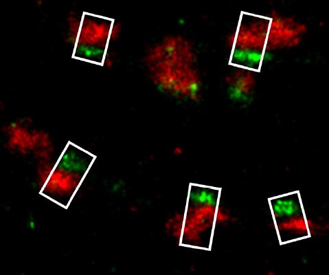
Super-Resolution Microscopy Reveals Presynaptic Localization of the ALS/FTD Related Protein FUS in Hippocampal Neurons
M. Schoen, J. M. Reichel, M. Demestre, S. Putz, D. Deshpande, C. Proepper, S. Liebau, M. J. Schmeisser, A. C. Ludolph, J. Michaelis and T. M. Boeckers
Fused in Sarcoma (FUS) is a multifunctional RNA-/DNA-binding protein, which is involved in the pathogenesis of the neurodegenerative disorders amyotrophic lateral sclerosis (ALS) and frontotemporal dementia (FTD). A common hallmark of these disorders is the abnormal accumulation of mutated FUS protein in the cytoplasm. Under normal conditions FUS is confined to the nuclear compartment, in neurons, however, additional somatodendritic localization can be observed. In this study, we carefully analyzed the subcellular localization of endogenous FUS at synaptic sites of hippocampal neurons which are among the most affected cell types in FTD with FUS pathology. We could confirm a strong nuclear localization of FUS as well as its prominent and widespread neuronal expression throughout the adult and developing rat brain, particularly in the hippocampus, the cerebellum and the outer layers of the cortex. Intriguingly, FUS was also consistently observed at synaptic sites as detected by neuronal subcellular fractionation as well as by immunolabeling. To define a pre- and/or postsynaptic localization of FUS, we employed super-resolution fluorescence localization microscopy. FUS was found to be localized within the axon terminal in close proximity to the presynaptic vesicle protein Synaptophysin1 and adjacent to the active zone protein Bassoon, but well separated from the postsynaptic protein PSD-95. Having shown the presynaptic localization of FUS in the nervous system, a novel extranuclear role of FUS at neuronal contact sites has to be considered. Since there is growing evidence that local presynaptic translation might also be an important mechanism for plasticity, FUS – like the fragile X mental retardation protein FMRP – might act as one of the presynaptic RNA-binding proteins regulating this machinery. Our observation of presynaptic FUS should foster further investigations to determine its role in neurodegenerative diseases such as ALS and FTD.
Front. Cell. Neurosci., 2016, 9:496.
DOI: 10.3389/fncel.2015.00496