Publications 2014
Lipid membrandes and single ion channel recording for the advanced physics laboratora
Y. Klapper, K. Nienhaus, C. Röcker, G.U. Nienhaus
We present an easy-to-handle, low-cost, and reliable setup to study various physical phenomena on a nanometer-thin lipid bilayer using the so-called black lipid membrane technique. The apparatus allows us to precisely measure optical and electrical properties of free-standing lipid membranes, to study the formation of single ion channels, and to gain detailed information on the ion conduction properties of these channels using statistical physics and autocorrelation analysis. The experiments are well suited as part of an advanced physics or biophysics laboratory course; they interconnect physics, chemistry, and biology and will be appealing to students of the natural sciences who are interested in quantitative experimentation.
Am.J.Phys. 82 (2014) 502-509
DOI: 10.1119/1.4849815
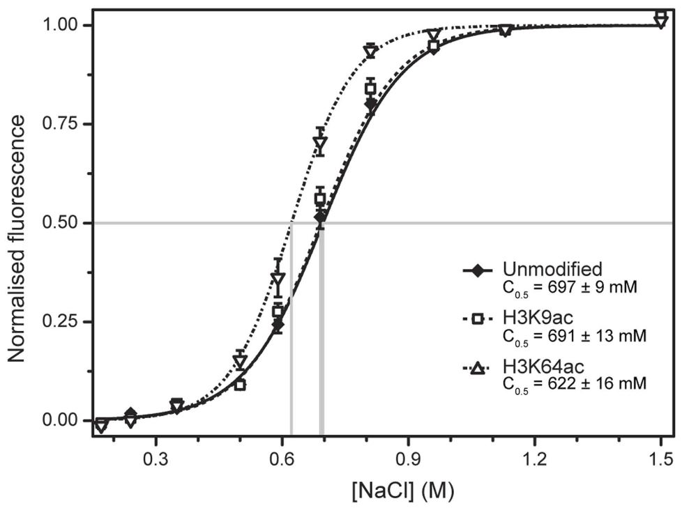
Acetylation of histone H3 at lysine 64 regulates nucleosome dynamics and facilitates transcription
V. di Cerbo, F. Mohn, D.P. Ryan, E. Montellier, S. Kacem, P. Tropberger, E. Kallis, M. Holzner, L. Hoerner, A Feldmann, F.M. Richter, A.J. Bannister, G. Mittler, J. Michaelis, S. Khochbin, R. Feil, D. Schuebeler, T. Owen-Hughes, S. Daujat, R. Schneider
Post-translational modifications of proteins have emerged as a major mechanism for regulating gene expression. However, our understanding of how histone modifications directly affect chromatin function remains limited. In this study, we investigate acetylation of histone H3 at lysine 64 (H3K64ac), a previously uncharacterized acetylation on the lateral surface of the histone octamer. We show that H3K64ac regulates nucleosome stability and facilitates nucleosome eviction and hence gene expression in vivo. In line with this, we demonstrate that H3K64ac is enriched in vivo at the transcriptional start sites of active genes and it defines transcriptionally active chromatin. Moreover, we find that the p300 co-activator acetylates H3K64, and consistent with a transcriptional activation function, H3K64ac opposes its repressive counterpart H3K64me3. Our findings reveal an important role for a histone modification within the nucleosome core as a regulator of chromatin function and they demonstrate that lateral surface modifications can define functionally opposing chromatin states.
eLIFE (2014);3:e01632
DOI: 10.7554/eLIFE.01632
Simultaneous dual-color 3D STED microscopy
C. Osseforth, J.R. Moffitt, L. Schermelleh and J. Michaelis
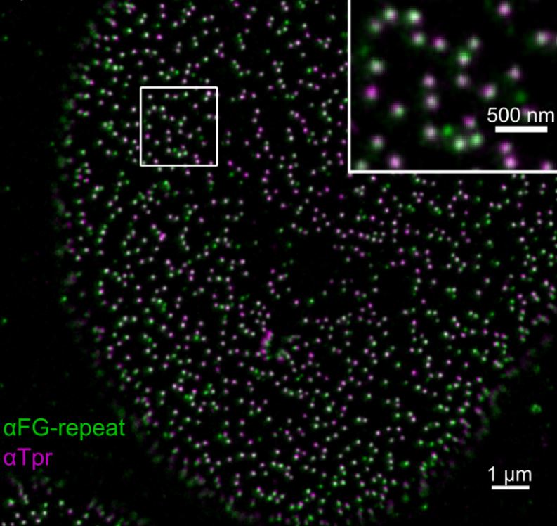
We describe the design and implementation of a stimulated emission depletion (STED) microscope which allows simultaneous three-dimensional super-resolution imaging in two colors. A super-continuum laser source is used to provide all spectral bands necessary for excitation and efficient depletion to achieve a lateral and axial resolution of ~35 nm and ~90 nm respectively. We characterize the systems' performance by imaging colloidal particles and single fluorescent molecules. Its biological applicability is demonstrated by dual-color imaging of nuclear pore complexes and of DNA replication sites in mammalian cells.
Optics Express (2014) 22, 7028-7039
DOI: 10.1364/OE.22.007028
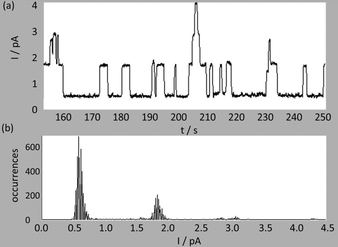
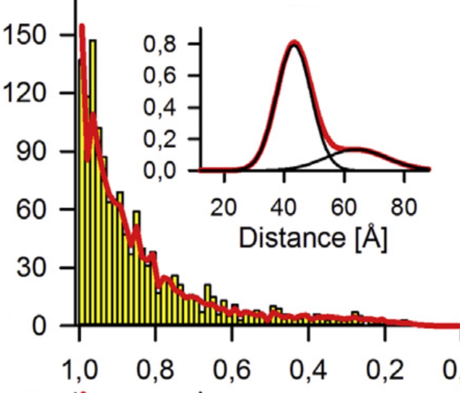
Nucleotides and Substrates Trigger the Dynamics of the Toc34 GTPase Homodimer Involved in Chloroplast Preprotein Translocation
C. Lumme, H. Altan-Martin, R. Dastvan, M. S. Sommer, M. Oreb, D. Schuetz, B. Hellenkamp, O. Mirus, J. Kretschmer, S. Lyubenova, W. Kügel, J.P. Medelnik, M. Dehmer, J. Michaelis, T. F. Prisner, T. Hugel and E. Schleiff
GTPases are molecular switches that control numerous crucial cellular processes. Unlike bona fide GTPases, which are regulated by intramolecular structural transitions, the less well studied GAD-GTPases are activated by nucleotide-dependent dimerization. A member of this family is the translocase of the outer envelope membrane of chloroplast Toc34 involved in regulation of preprotein import. The GTPase cycle of Toc34 is considered a major circuit of translocation regulation. Contrary to expectations, previous studies yielded only marginal structural changes of dimeric Toc34 in response to different nucleotide loads. Referencing PELDOR and FRET single-molecule and bulk experiments, we describe a nucleotide-dependent transition of the dimer flexibility from a tight GDP- to a flexible GTP-loaded state. Substrate binding induces an opening of the GDP-loaded dimer. Thus, the structural dynamics of bona fide GTPases induced by GTP hydrolysis is replaced by substrate-dependent dimer flexibility, which likely represents a general regulatory mode for dimerizing GTPases.
Structure (2014) 22, 526-538
DOI:10.1016/j.str.2014.02.004
Cyclophilin-Facilitated Membrane Trabslocation as Pharmacological Target to Prevent Intoxication of Mammalian Cells by Binary Clostridial Actin ADP-Ribosylated Toxins
K. Ernst, S. Langer, E. Kaiser, C. Osseforth, J. Michaelis, M.R. Popoff, C. Schwan, K. Aktories, V. Kahlert, M. Malesevic, C. Schiene-Fischer, H. Barth
Clostridiumbotulinum C2 toxin, Clostridiumperfringens iota toxin and Clostridiumdifficile CDT belong to the family of binary actin ADP-ribosylating toxins and are composed of a binding/translocation component and a separate enzyme component. The enzyme components ADP-ribosylate G-actin in the cytosol of target cells resulting in depolymerization of F-actin, cell rounding and cell death. The binding/translocation components bind to their cell receptors and form complexes with the respective enzyme components. After receptor-mediated endocytosis, the binding/translocation components form pores in membranes of acidified endosomes and the enzyme components translocate through these pores into the cytosol. This step is facilitated by the host cell chaperone heat shock protein 90 and peptidyl-prolyl cis/trans isomerases including cyclophilin A. Here, we demonstrate that a large isoform of cyclophilin A, the multi-domain enzyme cyclophilin 40 (Cyp40), binds to the enzyme components C2I, Ia and CDTa in vitro. Isothermal titration calorimetry revealed a direct binding to C2I with a calculated affinity of 101 nM and to Ia with an affinity of 1.01 μM. Closer investigation for the prototypic C2I revealed that binding to Cyp40 did not depend on its ADP-ribosyltransferase activity but was stronger for unfolded C2I. The interaction of C2I with Cyp40 was also demonstrated in lysates from C2-treated cells by pull-down. Treatment of cells with a non-immunosuppressive cyclosporine A derivative, which still binds to and inhibits the peptidyl-prolyl cis/trans isomerase activity of cyclophilins, protected cells from intoxication with C2, iota and CDT toxins, offering an attractive approach for development of novel therapeutic strategies against binary actin ADP-ribosylating toxins.
Journal of Molecular Biology (2015) 427, 1224-1238
DOI:10.1016/j.jmb.2014.07.013
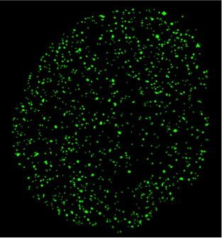
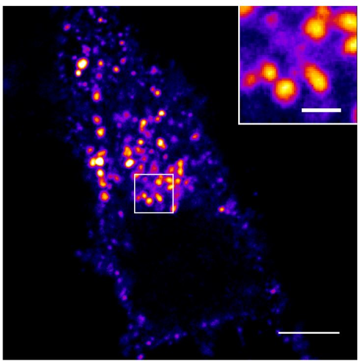
Spatial organization of RNA polymerase II inside a mammalian cell nucleus revealed by reflected light-sheet superresolution microscopy
Z.W. Zhao*, R. Roy*, J.C.M. Gebhardt*, D.M. Suter*, A.R. Chapman and X.S. Xie
Superresolution microscopy based on single-molecule centroid determination has been widely applied to cellular imaging in recent years. However, quantitative imaging of the mammalian nucleus has been challenging due to the lack of 3D optical sectioning methods for normal-sized cells, as well as the inability to accurately count the absolute copy numbers of biomolecules in highly dense structures. Here we report a reflected light-sheet superresolution microscopy method capable of imaging inside the mammalian nucleus with superior signal-to-background ratio as well as molecular counting with single-copy accuracy. Using reflected light-sheet superresolution microscopy, we probed the spatial organization of transcription by RNA polymerase II (RNAP II) molecules and quantified their global extent of clustering inside the mammalian nucleus. Spatiotemporal clustering analysis that leverages on the blinking photophysics of specific organic dyes showed that the majority (>70%) of the transcription foci originate from single RNAP II molecules, and no significant clustering between RNAP II molecules was detected within the length scale of the reported diameter of “transcription factories.” Colocalization measurements of RNAP II molecules equally labeled by two spectrally distinct dyes confirmed the primarily unclustered distribution, arguing against a prevalent existence of transcription factories in the mammalian nucleus as previously proposed. The methods developed in our study pave the way for quantitative mapping and stoichiometric characterization of key biomolecular species deep inside mammalian cells.
(* equal contribution)
Proc. Natl. Acad. Sci. (2014) 111, 681-686
DOI: 10.1073/pnas.1318496111