B09: Elucidating the pathomechanisms of compromised fracture healing in osteoporosis: Consequences of mitochondrial dysfunction
PIs: A. Ignatius, J. Riegger-Koch
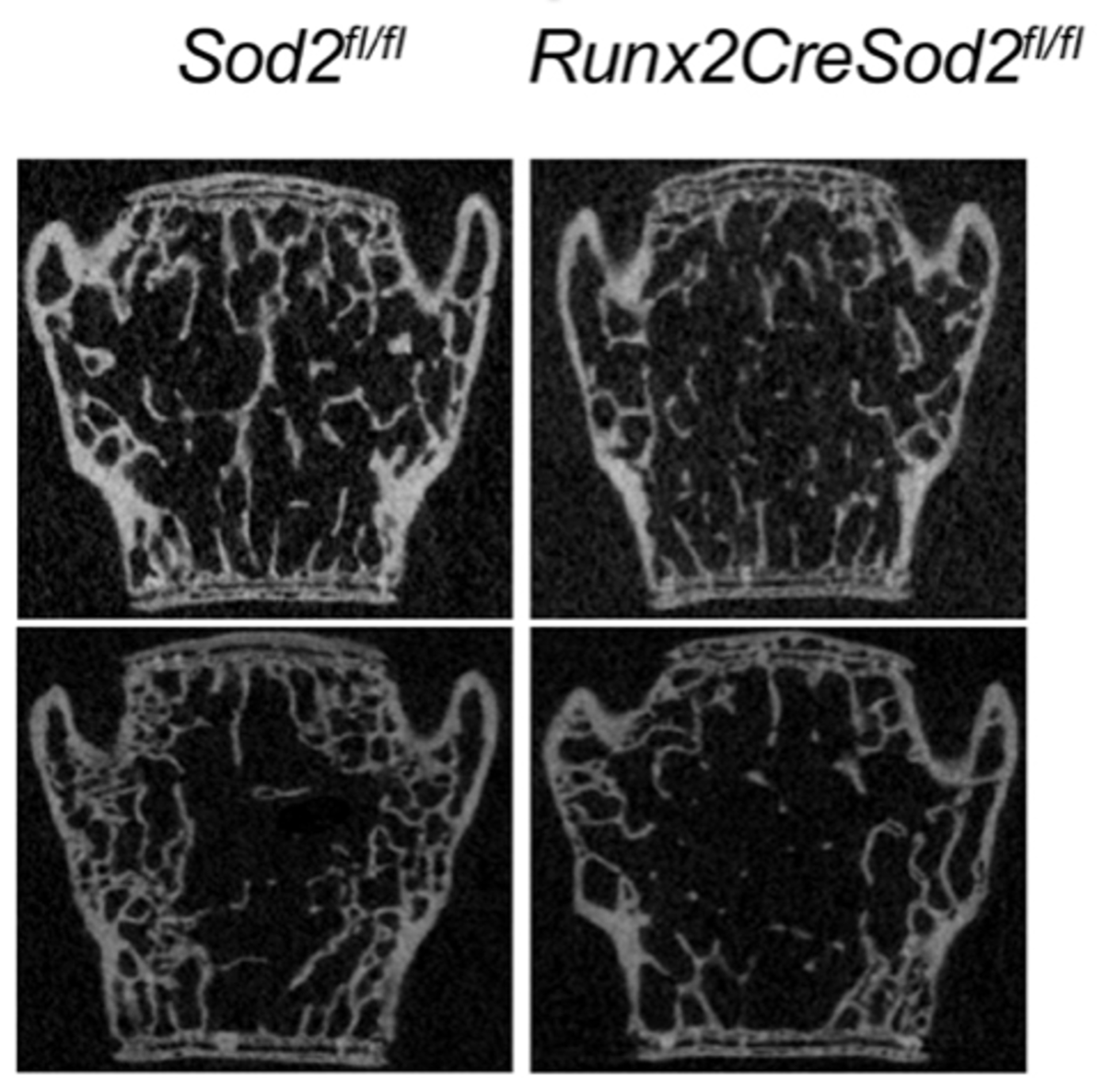
Osteoporotic fractures are frequently associated with healing complications, but the underlying mechanisms are still unclear. Previously, we demonstrated that mitochondrial (mt) ROS accumulation in osteoblasts disturbs osteogenic differentiation, pathways involved in mitophagy/autophagy, and cellular senescence, using a new mouse model with osteoblast lineage specific Sod2 deficiency, which phenocopies age-related osteoporosis. Therefore, we hypothesize that excessive mtROS production in osteoporotic bone is a key driver of compromised fracture healing by impairing intramembranous and endochondral bone formation. Combining bone healing experiments in mice with a Sod2 deletion specifically in osteoblast lineage cells or hypertrophic chondrocytes with comprehensive in vitro studies, we aim to elucidate how excessive mtROS affect osteoblast and chondrocyte function and fate during fracture healing. We will also study whether pharmacological stabilization of mt function ameliorates compromised fracture healing in mice with mtROS-induced osteoporosis as a potential therapeutic approach. Our results will help to better understand the role of oxidative stress in fracture healing and to develop novel therapies to improve bone healing in osteoporotic patients.
Projektleiterinnen
Prof. Dr. Anita Ignatius
Institut für Unfallchirurgische Forschung und Biomechanik
Zentrum für Traumforschung Ulm
Universitätsklinikum Ulm
Helmholtzstr. 14
89081 Ulm
Tel.: +49 731 500 55301
Fax: +49 731 500 55302
anita.ignatius(at)uni-ulm.de
www.biomechanics.de
Dr. biol. hum. Jana Riegger-Koch
Universitätsklinikum Ulm c/o RKU
Sektion Biochemie für Gelenks- und Bindegewebserkrankungen
Oberer Eselsberg 45
89081 Ulm
Tel.: +49 731 500 63288
E-mail: jana.riegger(at)uni-ulm.de