Overview
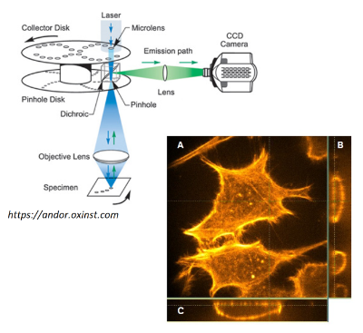
Spinning-disk confocal microscopy is applied using different fluorescence tagging strategies.
The actin cytoskeleton of fixed HeLa cells is stained by rhodamine phalloidin, which acts like a highly specific fluorescent antibody.
Intracellular calcium signals following receptor stimulation are studied using Fluo4, a cell permeant calcium indicator dye.
Dynamics in the mitochondrial network is studied using mt-Eos, a photoactivatable protein which carries a mitochondrial targeting signal.